The Extraordinary Science of How Humans Hear
From the folds of the outer ear to a living amplifier coiled inside the skull, hearing is a chain of mechanical marvels that turns a ripple of air into meaning in a fraction of a second.

A violinist draws her bow and, a few metres away, the air begins to shiver. The pressure changes are tiny, far smaller than the change you would feel climbing a flight of stairs. Yet a moment later two thousand people in a concert hall hear the note. Most of them could tell you whether it was a violin or a viola, whether it came from the left of the stage, and whether it was played with tenderness or menace.
All of that depends on structures smaller than a grain of rice, a few thousand specialised cells and a biological amplifier understood only within living memory.
The ear you can see
The outer ear, or pinna, looks like decoration, but its ridges and hollows do a job. As sound bounces around the folds, some frequencies are boosted and others dimmed, in a pattern that depends on whether the sound comes from above, below, in front or behind. The brain reads these subtle colourings as clues to where a sound is.
It must learn them. In 1998 Paul Hofman, Johannes Van Riswick and John Van Opstal, writing in Nature Neuroscience, fitted volunteers with moulds that reshaped their outer ears. At first they were very poor at judging how high a sound was, but they steadily became accurate again. When the moulds came off, they could still locate sounds with their own ears. The brain had kept both maps.
Three tiny bones
At the end of the ear canal sits the eardrum, a membrane that vibrates in time with the incoming sound. Behind it lie the three ossicles: the malleus (hammer), incus (anvil) and stapes (stirrup). The stapes is the smallest bone in the human body.
They solve an engineering problem. Sound travels easily through air but bounces off water, and the inner ear is filled with fluid. So the middle ear concentrates the energy. The vibrating part of the eardrum is roughly 13 to 20 times larger than the stapes footplate, by textbook estimates, and the bones add a little lever action. The same force pushed through a smaller area raises the pressure, much as a stiletto heel dents a floor that a flat shoe would not.

A piano rolled into a snail
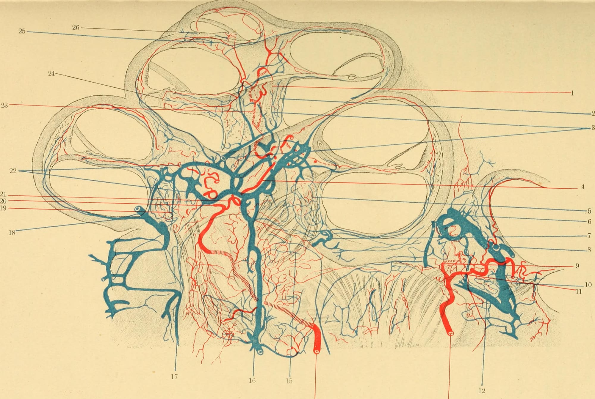
The stapes pushes like a piston against a small membrane-covered window into the cochlea. This is a fluid-filled spiral of about two and three-quarter turns which, if uncoiled, would measure about 35 millimetres. Along its length runs the basilar membrane, which supports the sensory cells.
How the cochlea separates pitches was worked out largely by the Hungarian-born physicist Georg von Békésy, who sprinkled the membrane with fine silver particles and watched them under flashing light. Sound, he found, sets up a travelling wave that ripples from the base of the cochlea towards its tip, peaking at a different place for each frequency. The stiff, narrow base responds to high notes, and the floppier far end responds to low ones. This orderly layout is called tonotopy, and it is preserved all the way up to the auditory cortex. Békésy received the 1961 Nobel Prize in Physiology or Medicine for the work. In effect, the cochlea is a keyboard, rolled up and read in reverse.
Healthy young ears span roughly 20 Hz to 20,000 Hz, about ten octaves. The eye, by comparison, sees less than one octave of light.
Hair cells, and the amplifier inside
The sensory work is done by hair cells, named for the tufts of stiff stereocilia on top. When the basilar membrane moves, the tufts bend, channels at their tips open, ions flood in, and movement becomes an electrical signal.
There are two kinds. A single row of inner hair cells, roughly 3,000 to 4,000 of them, often quoted as about 3,500, does most of the listening. Around 95% of the fibres of the auditory nerve connect to these cells. Three rows of outer hair cells, roughly 12,000 or more, do something stranger. They move.
In 1985 William Brownell and colleagues showed that isolated outer hair cells shorten and lengthen when a current passes through them. In 2000 Peter Dallos's group at Northwestern University identified the protein responsible and named it prestin, after the musical direction presto. Outer hair cells pump in time with the vibration, feeding energy back into the membrane and sharpening its tuning.
The ear is not a passive microphone. It is an instrument that plays along with the music it hears.
The size of the boost became clear in 2002, when Charles Liberman and colleagues bred mice without prestin. Their outer hair cells no longer moved, and their hearing became 40 to 60 decibels less sensitive. This is the cochlear amplifier. It is also what tends to fail first. A 2019 study of donated human ears found that people over 60 had lost on average 30 to 40% of their outer hair cells, while inner hair cell losses rarely exceeded 15%.
:::note The numbers
- About 3,500: inner hair cells in each human cochlea, in a single row
- 12,000+: outer hair cells, in three rows, acting as the cochlear amplifier
- About 95%: share of auditory nerve fibres that connect to inner hair cells
- 2¾ turns, about 35 mm: the coiled length of the cochlea
- 40–60 dB: the loss in sensitivity when mice lack prestin, the outer hair cells' motor protein :::
The ear that sings back
The amplifier has a strange side effect. In 1978 the British physicist David Kemp, at the Royal National Throat, Nose and Ear Hospital in London, placed a sensitive microphone in the ear canal and played brief clicks. After the expected echo from the middle ear came a faint, slower response from the cochlea itself, present in every normal ear he tested and absent in ears with cochlear deafness.
These otoacoustic emissions are sounds made by the ear as its outer hair cells work. Because they can be measured in seconds without any response from the listener, they made it practical to screen newborn babies, who obviously cannot press a button when they hear a beep. The Royal Society credits Kemp's discovery with helping to make possible the NHS newborn hearing screening programme.
Where hearing really happens
From the hair cells, signals travel up the auditory nerve through relay stations in the brainstem, where timing differences between the two ears are compared to within millionths of a second, and on to the auditory cortex. The ear analyses sound. Recognising a melody, a voice or a word is the brain's work.
Human cochlear hair cells do not grow back, which is why caring for your ears matters. If music has started to sound muffled, or you often ask people to repeat themselves, a hearing check through your GP or a local audiology service is simple, painless and worth having.
Sources & further reading
- NIDCD: How do we hear?
- Hofman, P. M., Van Riswick, J. G. & Van Opstal, A. J. (1998). Relearning sound localization with new ears. Nature Neuroscience
- BATOD: The inner ear (cochlear dimensions and hair cell numbers)
- Cochlea.eu: Inner hair cells, an overview
- HyperPhysics: von Békésy and the place theory
- Brownell, W. E. et al. (1985). Evoked mechanical responses of isolated cochlear outer hair cells. Science
- Zheng, J. et al. (2000). Prestin is the motor protein of cochlear outer hair cells. Nature
- Liberman, M. C. et al. (2002). Prestin is required for electromotility of the outer hair cell and for the cochlear amplifier. Nature
- Kemp, D. T. (1978). Stimulated acoustic emissions from within the human auditory system. JASA
- Wu, P. Z. et al. (2019). Primary neural degeneration in the human cochlea. Neuroscience
- Royal Society: Professor David Kemp FRS
Filed under Hearing & Sound. Spotted an error? Tell the editors.